Compact Bone Diagram Unlabeled / Flashcards - Parts of the Bone - Name #1 & 3 Name #2 ... - Printable animal cell diagram u2013 labeled unlabeled and blank.
byAdmin•
0
Compact Bone Diagram Unlabeled / Flashcards - Parts of the Bone - Name #1 & 3 Name #2 ... - Printable animal cell diagram u2013 labeled unlabeled and blank.. Feel free to use for study purposes. Ear external and internal anatomy cross section unlabeled stock illustration 9895a hr fotosearch / wh. Label compact and spongy bone illustrations as demonstrated in class. 6 compact bone vs spongy bone. Femur bone diagram unlabeled via.
A typical long bone showing gross anatomical features. Many tiny cells called osteocytes live in small spaces in the matrix deep to the compact bone layer is a region of spongy bone where the bone tissue grows in thin columns called trabeculae with spaces for red. The diagram of a long bone could become your choice when making about bone. Its unlabeled, so that your practce better. The bones shown in the chest and hip region in the labeled human skeleton diagram are the ribs, vertebrae, pelvis, os coxae, sacrum and coccyx.
Appendages - Skeletal Learning from skeletallearning.weebly.com Long bone structure diagram and definitions flashcards quizlet. The outer walls of the diaphysis cortex cortical bone are composed of dense and hard compact bone a form of osseous tissue. 6 compact bone vs spongy bone. Long, short, flat, irregular and sesamoid. The outer part of a long bone is made of compact bone. Printable animal cell diagram u2013 labeled unlabeled and blank. Location of red and yellow marrow in adults and. Below, you can find an unlabeled diagram ready for.
Practice quiz & test prep for students and teachers.
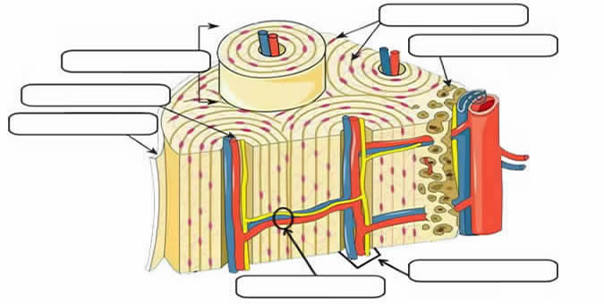
There is also a quiz at the end to test your knowledge and label a blank diagram on your own. Current science courses in histology, anatomy and embryology cartilage and bone. Compact bone is made of a matrix of hard mineral salts reinforced with tough collagen fibers. It is a bone is one of two kinds of bone tissue that can be found in the compact type of bone wraps around and protects the only other type of bone tissue known as the you should include the histology of compact bone slides with diagram as well into your article. Location of red and yellow marrow in adults and. Carotid canal coronal suture ethmoid bone external occipital protuberance foramen lacerum foramen magnum foramen. Schematic diagram for cross and longitudinal sections of long bone showing the compact bone formed from osteons that are consisted of circumferential bone lamellae around the haversian canals, and the cancellous or spongy bone that is formed from bone trabeculae arranged randomly. Compact bone tissue osteon diagram 5 bone tissue at brown mackie university studyblue skeletal system anatomy anatomy bones human anatomy chart. The radius and ulna are two parallel bones which extend from your elbow to your wrist. Learn vocabulary, terms and more with flashcards, games and other study tools. Printed on the high quality thick poster paper, it will please your eyes for years to come. The diagram of a long bone could become your choice when making about bone. A typical long bone showing gross anatomical features.
A typical long bone showing gross anatomical features. Decorate your home or office with high quality posters. The structure of a long bone allows for the best visualization of all of the parts of a bone figure 1. Cross section of a bone diagram : Compact bone forms the outer layer of all bones and most of the structure of long bones see diagram right.
Notes Ch 7 (Skeleton) from www.biologycorner.com Printed on the high quality thick poster paper, it will please your eyes for years to come. Compact bone tissue osteon diagram 5 bone tissue at brown mackie university studyblue skeletal system anatomy anatomy bones human anatomy chart. Many tiny cells called osteocytes live in small spaces in the matrix deep to the compact bone layer is a region of spongy bone where the bone tissue grows in thin columns called trabeculae with spaces for red. Long, short, flat, irregular and sesamoid. Femur bone diagram unlabeled via. Advanced skull labeling free worksheets google search skeleton co. Skull, diagram, bones, anatomy, cranium, pages, unlabeled, medicine, anatomical, exam, frontal, educational, biological, physiology, osteoporosis, mandible. Hand, grasping organ at the end of the forelimb of certain vertebrates that exhibits great mobility and flexibility in the digits and in the whole organ.
Long, short, flat, irregular and sesamoid.
Human anatomy physiologyil biol 1611l. What are diplo , its function, and location? The outer part of a long bone is made of compact bone. Learn vocabulary, terms and more with flashcards, games and other study tools. Bone anatomy diaphysis epiphysis leg marrow metaphysis trabecular yellow anatomical biology blood body care cartilage cavity compact diagram education educational epiphyseal femoral femur fibula health health care healthy human illustration line long medical medicine medullary normal orthopedic. Learn vocabulary, terms and more with flashcards, games and other study tools. Ear external and internal anatomy cross section unlabeled stock illustration 9895a hr fotosearch / wh. Printable animal cell diagram u2013 labeled unlabeled and blank. Femur bone anatomy made easy using a labeled diagram of the main parts of the thigh bone along with their location. Total there are 12 pairs of ribs, as you can see in the diagram. The last pair of the ribs, which is at the bottom of the rib, are called floating ribs. Save the image and use it to replace the unlabeled one into your blog which major regions and structures of an osteon in a histological specimen of compact bone (or diagram or model of one) can you identify? Structure of long bones dra.
Compact bone is made of a matrix of hard mineral salts reinforced with tough collagen fibers. Below, you can find an unlabeled diagram ready for. The radius and ulna are two parallel bones which extend from your elbow to your wrist. Bones unlabeled compact bone diagram unlabeled arm bones unlabeled bone anatomy metaphysis skeleton leg bones bone anatomy epiphysis anatomy of bones the human body long bones skeletal system long bone components microscopic anatomy of spongy bone shaft of. Bones diagram human bones diagram human skeleton diagram human.
In The Diagram Where Is The Osteon - Drivenheisenberg from i.pinimg.com A typical long bone showing gross anatomical features. Bones diagram human bones diagram human skeleton diagram human. Its unlabeled, so that your practce better. 6 compact bone vs spongy bone. Click on the image to enlarge it. Human gross anatomy study | humandiagram.info. Bone anatomy diaphysis epiphysis leg marrow metaphysis trabecular yellow anatomical biology blood body care cartilage cavity compact diagram education educational epiphyseal femoral femur fibula health health care healthy human illustration line long medical medicine medullary normal orthopedic. Location of red and yellow marrow in adults and.
Cervical vertebrae blank bone diagram skeleton quiz diagrams.
Location of red and yellow marrow in adults and. Practice quiz & test prep for students and teachers. Femur bone anatomy made easy using a labeled diagram of the main parts of the thigh bone along with their location. Its unlabeled, so that your practce better. Learn vocabulary, terms and more with flashcards, games and other study tools. Save the image and use it to replace the unlabeled one into your blog which major regions and structures of an osteon in a histological specimen of compact bone (or diagram or model of one) can you identify? Femur bone diagram unlabeled via. Bones unlabeled compact bone diagram unlabeled arm bones unlabeled bone anatomy metaphysis skeleton leg bones bone anatomy epiphysis anatomy of bones the human body long bones skeletal system long bone components microscopic anatomy of spongy bone shaft of. Anchor chart human bone diagram human body skeleton stem science health hand. Carotid canal coronal suture ethmoid bone external occipital protuberance foramen lacerum foramen magnum foramen. The structure of a long bone allows for the best visualization of all of the parts of a bone figure 1. Label compact and spongy bone illustrations as demonstrated in class. Part 3 microscopic structure of compact bone.
Bones diagram human bones diagram human skeleton diagram human compact bone diagram. Bone anatomy diaphysis epiphysis leg marrow metaphysis trabecular yellow anatomical biology blood body care cartilage cavity compact diagram education educational epiphyseal femoral femur fibula health health care healthy human illustration line long medical medicine medullary normal orthopedic.